Posterior segment shape in myopia: observations from the adult anisomyopia study
Although myopia is known to be associated with increased axial length of the eyeball, it remains unclear to what extent the transverse dimensions also change. Understanding how the posterior segment remodels is essential for explaining myopia development and for designing effective control strategies. The study conducted by Logan, Gilmartin, and colleagues (IOVS, 2004) provides relevant data by comparing ocular shape in isomyopic and anisomyopic individuals from two distinct ethnic populations.
Context and methodology
The study included 56 young adults, divided into four groups: isomyopes and anisomyopes, both Caucasian and Taiwanese-Chinese. Anisomyopic participants presented a minimum interocular difference of 2 D, which allows a controlled analysis of shape differences between two eyes exposed to the same genetic and environmental influences.
The reconstruction of the posterior retinal contour was achieved by integrating:
- central and peripheral refraction measured up to ±35°
- axial biometric data obtained through A-scan
- corneal curvature values
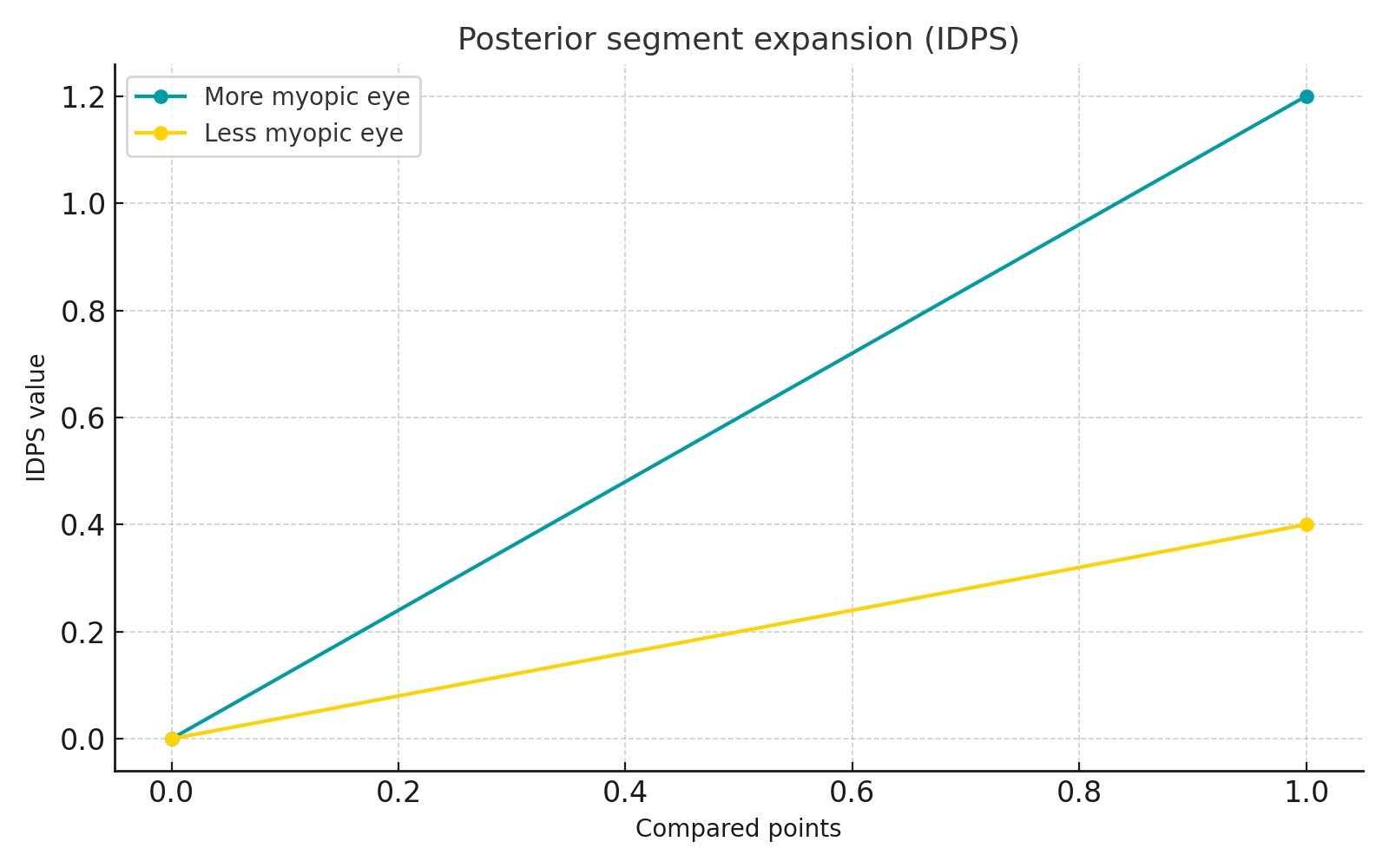
An optical algorithm generated 2D coordinates of the retinal surface based on ray modelling in the sagittal and tangential planes. Differences between the more myopic and the less myopic eye were quantified using the posterior distance elongation index (IDPS), which reflects the degree of posterior segment expansion.

Results
Pronounced axial growth and prolate contour
In both ethnic groups, the more myopic eyes showed greater axial length and a more prolate posterior segment shape. Expansion was primarily axial and exceeded the transverse enlargement, confirming that myopia is structurally characterised by an anteroposterior elongation.
Ethnic differences in expansion distribution
The analysis of retinal contours revealed clear differences between the two populations:
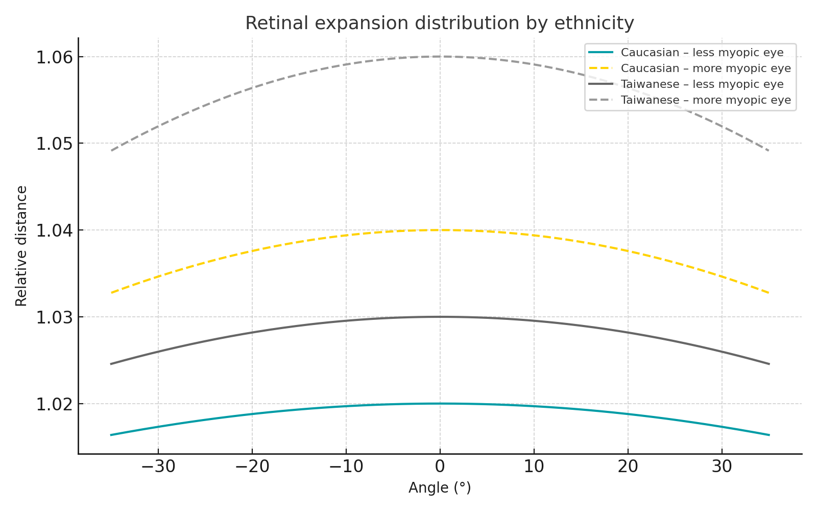
- in Caucasian subjects, expansion was asymmetric and more pronounced nasally
- in Taiwanese-Chinese subjects, expansion was greater and more symmetrical, involving both nasal and temporal sectors
This behaviour suggests biomechanical or histological particularities of the sclera between the two ethnicities, with potential implications for the higher susceptibility to myopia in Asian populations.
Relationship between transverse diameter and axial length
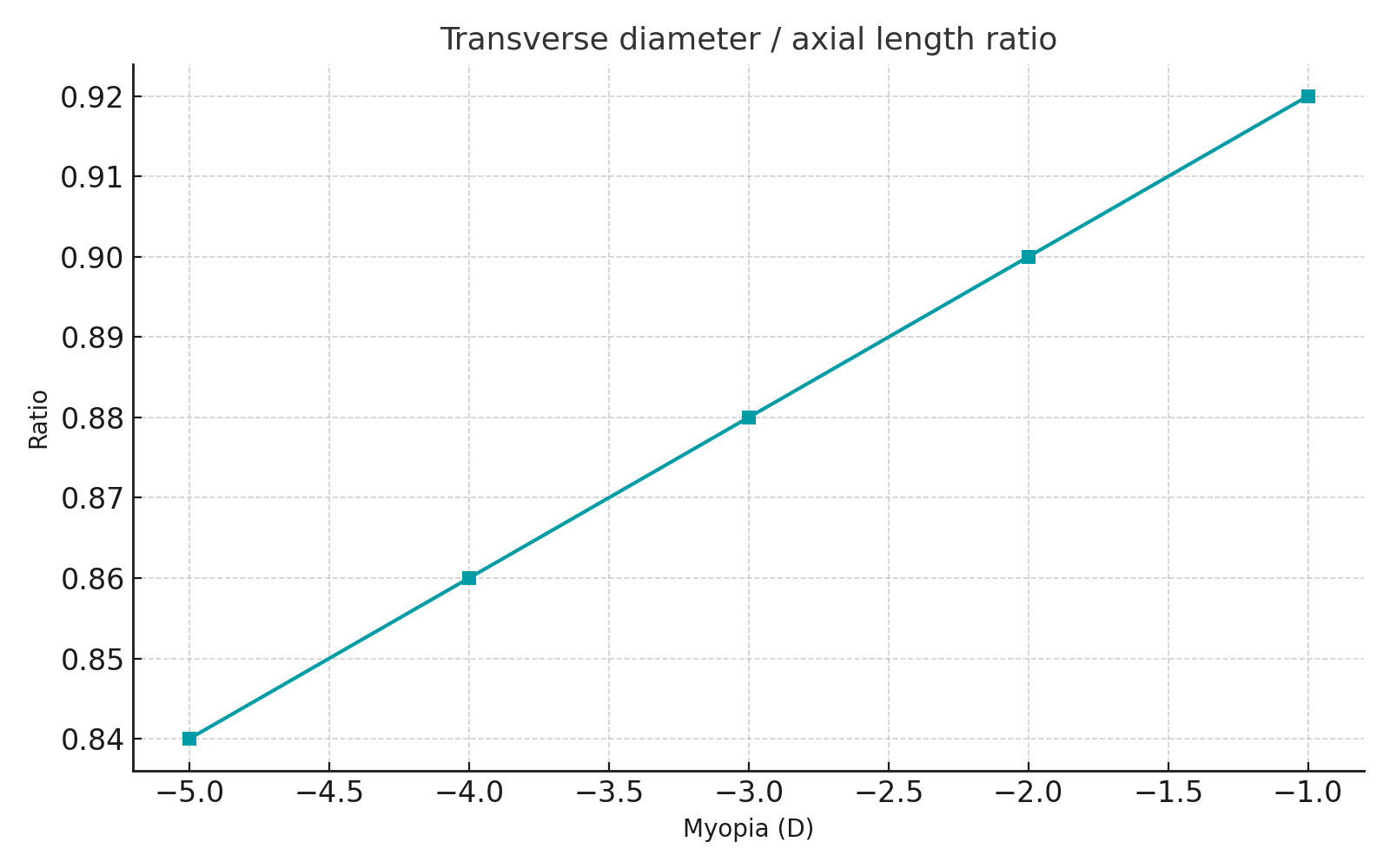
In the Taiwanese-Chinese group, the ratio between transverse diameter and axial length decreased with increasing myopia, indicating predominantly axial and consistent elongation. In contrast, no significant statistical relationship was found in the Caucasian group.
Changes in parapapillary crescent size
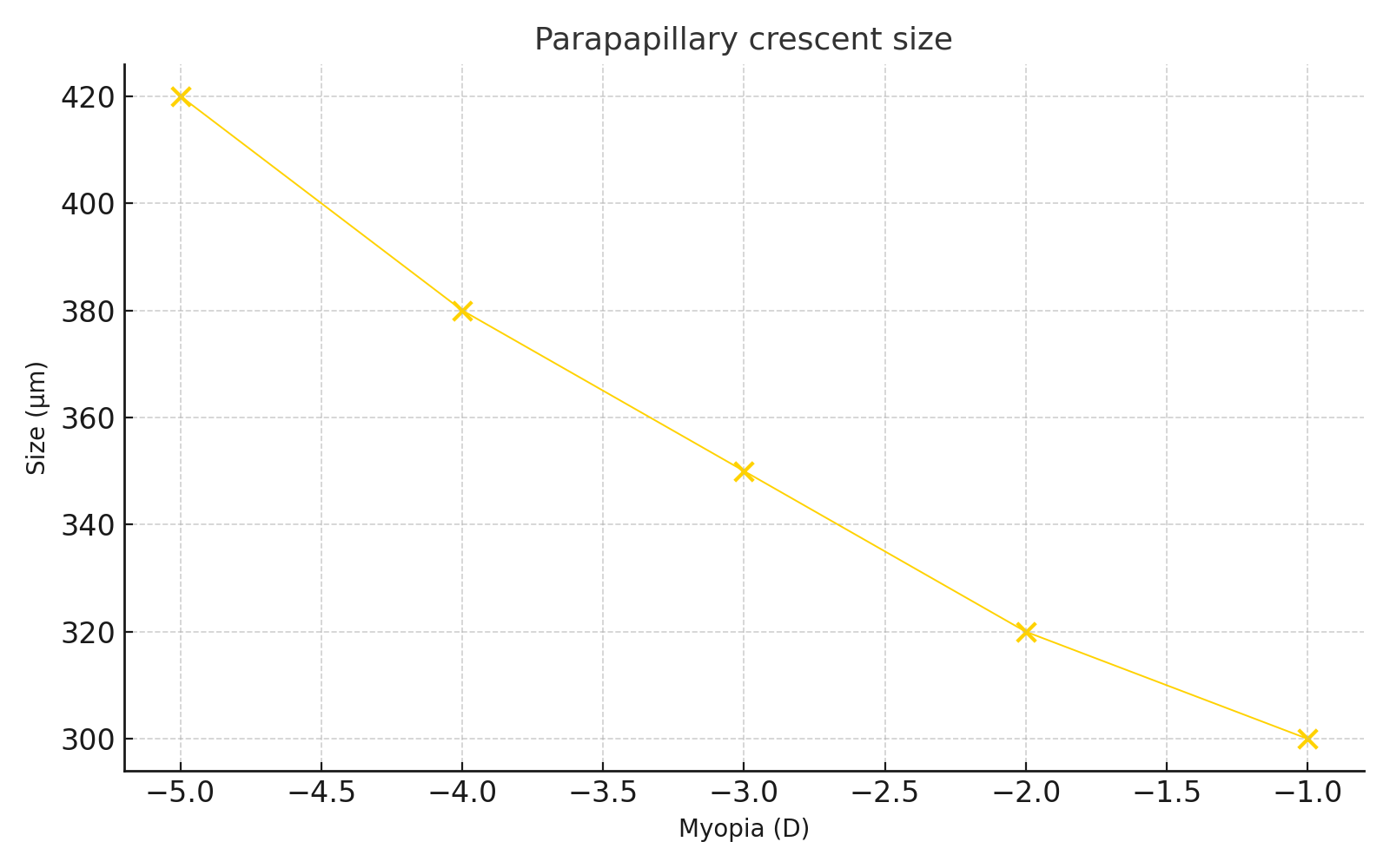
The study also evaluated the parapapillary crescent, a marker of retinal stretching:
- in Taiwanese-Chinese subjects, the crescent increased with the severity of myopia
- in Caucasian subjects, this relationship was not confirmed
Interpretation and analysis
The results indicate that posterior segment remodelling in myopia is not uniform but the result of a regionalised process in which the posterior sclera is more susceptible to stretching. The study suggests:
- a local control of retinal growth influenced by peripheral defocus
- structural differences between ethnicities, possibly related to scleral thickness or choroidal response
- a correspondence between the prolate shape and animal-model findings, which show more intense scleral–choroidal changes at the posterior pole
These observations support the hypothesis that the level and distribution of peripheral defocus influence ocular remodelling, with significant individual and ethnic variations.
Practical implications
For optometrists, ophthalmologists, and lens specialists, the study’s conclusions are useful for:
- interpreting myopia progression based on complete biometric data, not only axial length
- monitoring parapapillary crescent size as an indicator of structural stretching
- tailoring myopia-control strategies according to ethnicity
- validating optical approaches that modify peripheral defocus to reduce axial elongation signals
Conclusions
The study demonstrates that myopia is associated with axial elongation and with a prolate transformation of the posterior segment, more pronounced and more symmetrical in Taiwanese-Chinese subjects. Understanding these mechanisms is essential for developing effective optical and therapeutic solutions for myopia-control interventions.
Primary source: Logan N.S., Gilmartin B., Wildsoet C.F., Dunne M.C.M.
Posterior retinal contour in adult anisomyopia Investigative Ophthalmology & Visual Science (IOVS), vol. 45, no. 6, 2004, pp. 2152–2162.