Lenses for myopia management: the influence of peripheral defocus and retinal asymmetries on axial growth
Myopia is one of the major current challenges in the field of vision, not only because of its increasing incidence but also due to its long-term consequences, such as the elevated risk of glaucoma, macular degeneration, or retinal detachment. In recent years, research has focused on the role of the peripheral retina in controlling ocular growth, suggesting that the way peripheral images are focused can stimulate or inhibit axial elongation.
This principle forms the basis of modern lenses for myopia management, which aim to modify the distribution of peripheral defocus to reduce the eye’s growth stimulus. The clinical study analyzed provides a detailed perspective on the differences between stable and progressive myopia, highlighting key elements that must be considered in the design of these lenses.
Theoretical background: peripheral image formation in myopia
In the myopic eye, images of objects are formed in front of the retina. Standard correction with negative lenses restores central focus, but the peripheral retina is usually left in a state of hyperopic defocus. This condition creates a visual signal interpreted by the emmetropization system as a “need for elongation,” which may accelerate myopia progression.
Numerous studies on animal models have confirmed that:
- peripheral hyperopic defocus induces axial growth,
- myopic defocus has the opposite effect, inhibiting axial elongation of the eyeball,
- the peripheral retina can guide ocular growth even in the absence of central vision, underscoring its essential role.
Consequently, lenses for myopia management attempt to reverse this situation by introducing controlled peripheral myopic defocus.
Study methodology: analysis of peripheral refraction and retinal contour
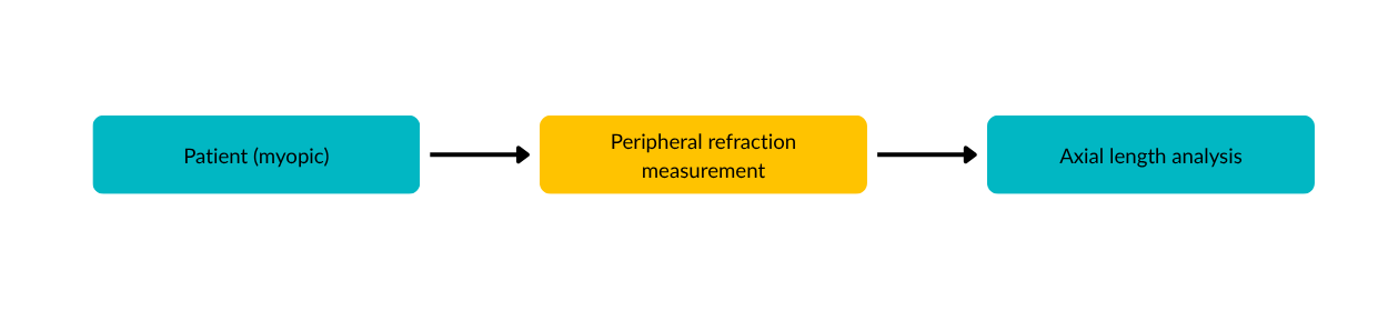
The study included 62 young Caucasian myopic eyes, divided into two groups:
- stable myopia (NP) – no significant refractive changes for at least two years;
- progressive myopia (P) – progression ≥ –0.50 D in the last year.
The groups were balanced regarding:
- mean age (~22 years),
- central refraction,
- axial length.
Detailed measurements were taken of:
- peripheral refraction on the horizontal meridian,
- peripheral astigmatic components (tangential and sagittal),
- eccentric axial length, used to reconstruct the retinal contour.
Results: fundamental differences between stable and progressive myopia
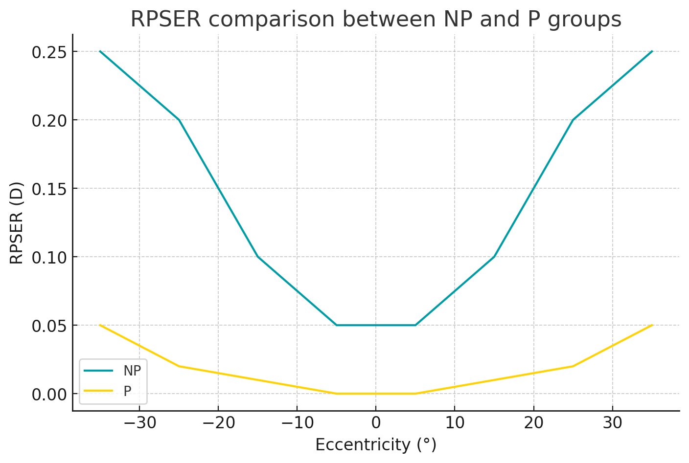
Peripheral equivalent refraction (RPSER)
The analysis of RPSER showed significant differences especially in the nasal hemifield:
- the P group presented values close to emmetropia,
- the NP group presented slightly myopic values.
This suggests that analyzing only the equivalent refraction is insufficient to explain axial growth behavior.
Positions of astigmatic foci (FT’ and FS’)
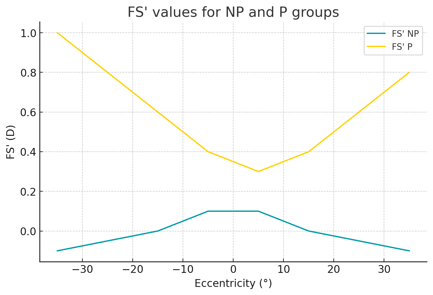
The most important differences were observed in sagittal focus (FS’) in the nasal hemifield:
- the P group showed significant peripheral sagittal hyperopic defocus (p < 0.001),
- the NP group showed myopic or nearly neutral values.
This peripheral sagittal hyperopia appears to be a critical stimulus for continued axial elongation.
Retinal contour: an important structural difference
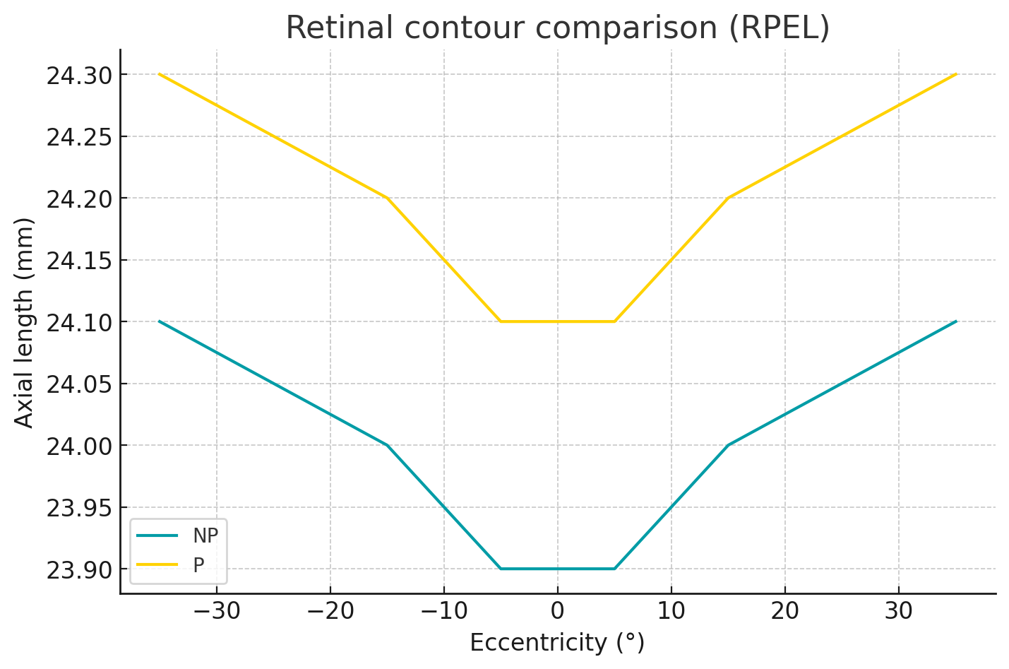
Measurements of eccentric axial length indicated:
- a steeper nasal retina in progressive myopes,
- a statistically significant difference (p = 0.001).
This anatomical configuration may amplify the effect of peripheral hyperopic defocus, creating a cycle that sustains myopia progression.
There is a strong inverse correlation (r ≈ –0.6 to –0.78) between:
• the degree of retinal curvature (RPEL),
• the tendency toward peripheral hyperopic defocus.
Analysis: the role of the nasal hemiretina in controlling ocular growth
A key finding of the study is the asymmetry between the nasal and temporal retina:
- marked differences between the P and NP groups appear predominantly nasally,
- the nasal retina may play a more important role in emmetropization mechanisms,
- this sector is exposed to a wider visual field and may be more sensitive to defocus.
Thus, lens designs that introduce differentiated myopic defocus, with increased emphasis in the nasal area, may be more effective in slowing myopia progression.
Implications for lens design for myopia management
Based on the results, effective myopia-control lenses should consider:
- Full correction of central refraction – ensures foveal image clarity and visual comfort.
- Stable peripheral myopic defocus – reduces the stimulus for axial elongation.
- Stronger positive power in the nasal area – addresses differences in retinal sensitivity and structure.
- Control of sagittal focus position, not only spherical equivalent – FS’ plays a critical role in the visual growth signal.
Such approaches align with both ocular growth theories and clinical study findings.
Conclusions
The analyzed study offers a detailed and compelling perspective on how:
- peripheral defocus,
- astigmatic focus position,
- retinal contour,
- nasal–temporal asymmetry,
influence myopia progression.
Progressive myopes display significant peripheral sagittal hyperopic defocus and a steeper nasal retina—factors that jointly contribute to stimulating axial growth.
These findings highlight the need to develop lenses that integrate intelligently distributed myopic defocus, adapted to retinal anatomical differences and the visual mechanisms involved in regulating ocular growth.
Main source: Donovan L., Sankaridurg P., Ho A., Chen X., Smith E.L., Holden B.A. Myopia progression in children wearing single‐vision spectacles: a meta‐analysis, Optometry and Vision Science, 2012, Vol. 89(1), pp. 27–32. Source: PubMed (DOI: 10.1097/OPX.0b013e3182357f79)