Predisposition for increased astigmatism in children using myopia management solutions: clinical study
Myopia progression control in children is a major priority for eye-care professionals in optometry and ophthalmology. In addition to axial elongation, changes in peripheral refraction—particularly the development of hyperopic defocus and peripheral astigmatic components—are considered important factors in ocular growth mechanisms.
With the growing use of modern myopia control solutions (soft contact lenses with peripheral defocus, orthokeratology, and ophthalmic lenses with multiple defocus zones), it is essential to understand how these interventions may influence peripheral astigmatism and, consequently, the risk of myopia progression.
This clinical review synthesizes data reported by Faria-Ribeiro et al. (2013), adapted to the pediatric population, to identify the predisposition toward increased astigmatism in children using myopia management solutions.
Background and methodology
The reference study analyzed differences in peripheral refraction and retinal contour between two groups of young myopes: one group with stable myopia and one with progressing myopia. Although participants were 20–22 years old, the physiological mechanisms identified are the same in children and are relevant for assessing progression risk in the pediatric population.
The authors evaluated:
- peripheral refraction (spherical equivalent and astigmatic FT and FS components),
- central and peripheral axial length,
- retinal shape in nasal and temporal hemifields.
Data were obtained using open-field autorefraction and optical biometry at eccentricities up to 30–35°.
In this article, interpretation refers to children using myopia management approaches such as peripheral-defocus soft contact lenses, orthokeratology lenses, or spectacle lenses designed for myopia control.
Results on peripheral refraction
The original study reports significant differences between eyes with stable and progressive myopia, particularly in the nasal retina, a visual field region highly sensitive to peripheral defocus.
Key observations:
- Children/young individuals with progressive myopia show more pronounced hyperopic defocus in the nasal periphery.
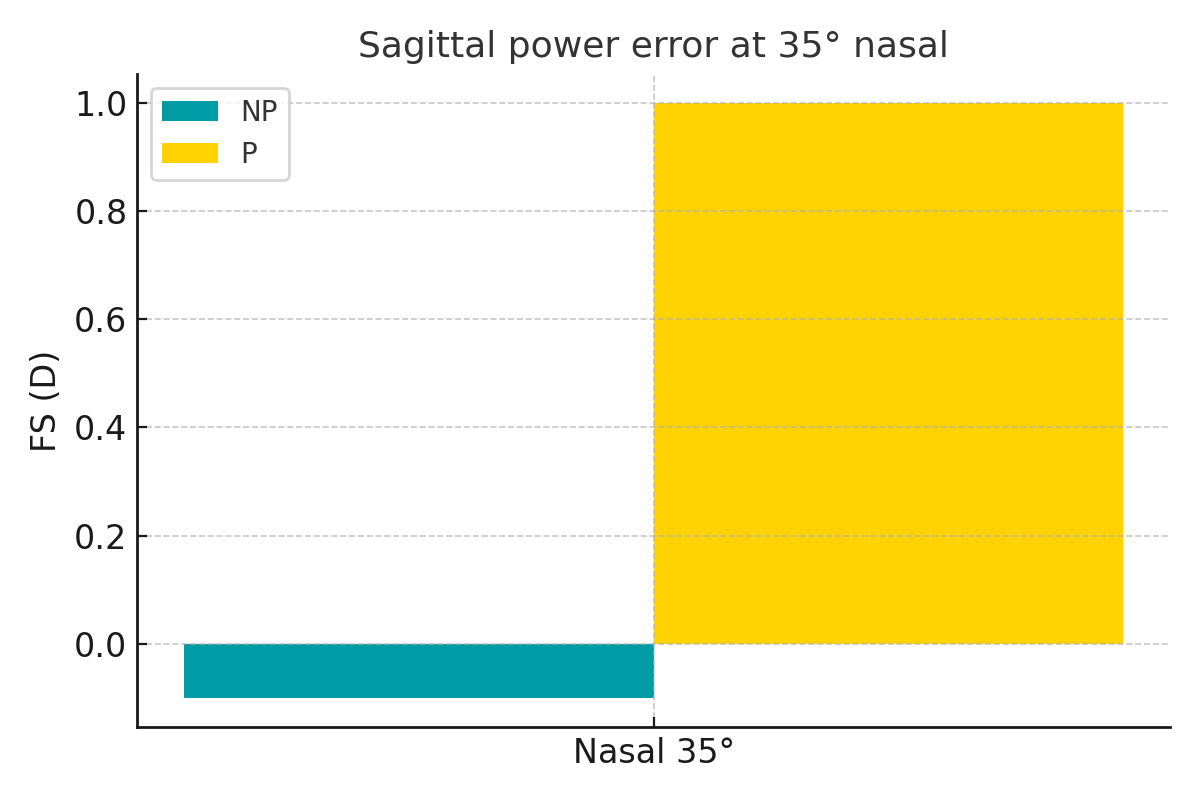
- The sagittal astigmatic component (FS) is primarily responsible for peripheral hyperopia: at 35° nasal, the progressing group showed a mean FS of +1.00 ± 0.83 D, compared to –0.10 ± 0.98 D in stable eyes.
- The tangential component (FT) remains closer to myopia in stable eyes, contributing to less hyperopic peripheral defocus.
Conclusion: hyperopic peripheral FS is associated with an increased risk of progression.
Retinal morphology and implications for axial growth
Retinal shape is an important indicator of predisposition to myopia progression. The study describes a steeper nasal retina in individuals with progressive myopia.
Relevant findings:
- Variations in peripheral axial length (RPEL) indicate a significantly steeper retina in the progression group (p = 0.001).
- There is a strong inverse correlation between RPEL and FS (r = –0.595 to –0.775).
→ The steeper the retina, the more hyperopic the sagittal defocus becomes.
Interpretation in the context of myopia management in children
Modern myopia control solutions aim to induce peripheral myopic defocus to interrupt the axial-length growth signal.
However, the study shows that:
- if peripheral sagittal refraction remains hyperopic, even when the peripheral spherical equivalent is emmetropic, the growth stimulus may persist;
- peripheral visual processing may interpret the position of astigmatic focal planes as a signal for elongation, regardless of spherical defocus;
- children with an already steeper nasal retinal profile are more likely to progress if the selected myopia management solution does not sufficiently reduce the FS component.
In practice, this means that some myopia control strategies may only partially reduce progression if they do not manage both peripheral astigmatic foci.
Clinical implications for practitioners
For children enrolled in a myopia control program, the following are recommended:
1. Peripheral refraction analysis along the horizontal meridian
- FS is a stronger progression predictor than spherical equivalent RPSER.
- Nasal hyperopic FS > +0.75 D suggests a high risk of progression.
2. Selection of optical treatment based on peripheral profile
- Myopia control contact lenses should be assessed according to their effectiveness in reducing FS.
- In orthokeratology, evaluating peripheral refraction after 1–2 weeks is essential.
3. Monitoring retinal morphology
- A steeper nasal retina indicates the need for stronger induced peripheral myopic defocus.
Conclusions
The analyzed data demonstrate that predisposition to increased peripheral astigmatism—particularly the elevation of the sagittal FS component—may explain why some children continue to progress even when using modern myopia control solutions.
Key Points:
- Hyperopic FS in the nasal periphery is a major risk signal for myopia progression.
- Optical myopia control solutions should be assessed not only for their influence on spherical defocus but also on peripheral astigmatism.
- The nasal retina shows increased sensitivity to defocus, and its shape directly affects treatment success.
These findings support a more personalized approach to myopia control in which peripheral refraction measurements and retinal shape become essential tools for effective therapy.
Main source: Faria-Ribeiro M., Navarro R., González-Méijome J.M. Peripheral refraction and retinal contour in stable and progressive myopic eyes. Investigative Ophthalmology & Visual Science, 2013.